In addition to the top three winners, the Nikon Small World Photomicrography Competition recognized 83 photos out of nearly 1,900 photo entries from 72 countries.
More info:nikonsmallworld.com|Instagram|twitter.com|Facebook
This post may includeaffiliate links.
Nikon Small World is very well known for being the leading forum where people celebrate the skill and beauty of photomicrography.Competition organizers shared that there are 4 factors to consider when evaluating submissions: originality, informational content, technical proficiency, and visual impact. To select the winners, judges looked at entries that came from all around the globe.
Nikon Small World is very well known for being the leading forum where people celebrate the skill and beauty of photomicrography.
Competition organizers shared that there are 4 factors to consider when evaluating submissions: originality, informational content, technical proficiency, and visual impact. To select the winners, judges looked at entries that came from all around the globe.
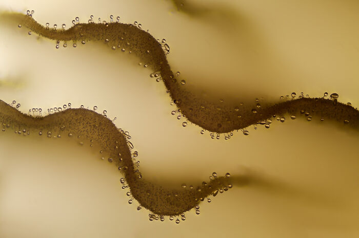
Universidad Complutense de MadridReal Sociedad Española de FísicaMadrid, Spain"Crystallized sugar syrup."
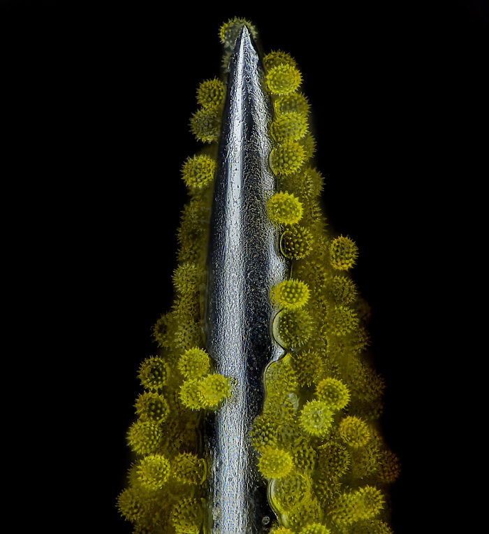
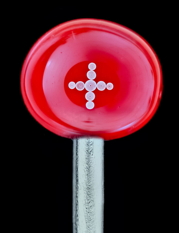
Medienbunker ProduktionBendorf, Rheinland Pfalz, Germany"Sunflower pollen on an acupuncture needle."
The University of GeorgiaAthens, Georgia, USA"Caffeine crystals."
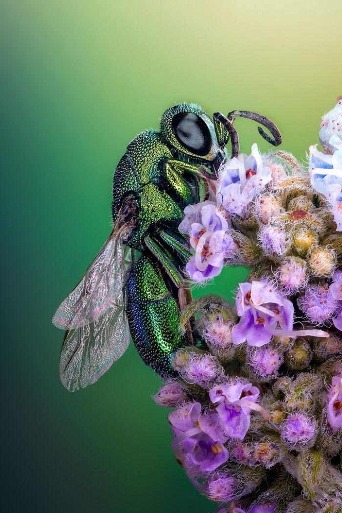
Tanta UniversityFaculty of ScienceDepartment of ZoologyTanta, Egypt"Cuckoo wasp standing on a flower."
The Nikon Small World competition was founded in 1974 and in 2011,Nikon Small World in Motionwas launched because of advancements in technology that made it possible to record videos or digital time-lapse photos through a microscope.The2023 video winnerswere announced on September 26th.
The Nikon Small World competition was founded in 1974 and in 2011,Nikon Small World in Motionwas launched because of advancements in technology that made it possible to record videos or digital time-lapse photos through a microscope.
The2023 video winnerswere announced on September 26th.
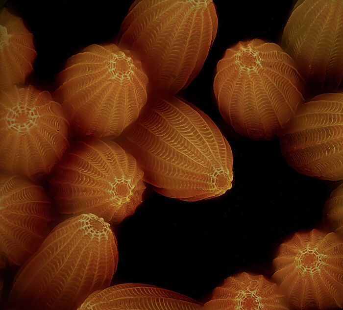
World Expo MuseumShanghai, China"Chinese moon moth (Actias ningpoana) wing scales."
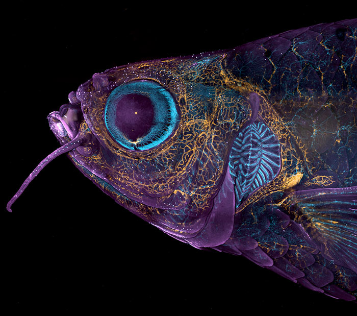
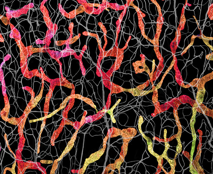
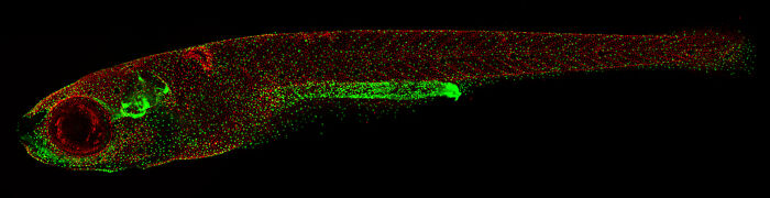
National Institutes of Health (NIH)Eunice Kennedy Shriver National Institute of Child Health and Human DevelopmentBethesda, Maryland, USA"Adult transgenic zebrafish head showing blood vessels (blue), lymphatic vessels (yellow), and the skin and scales (magenta)."
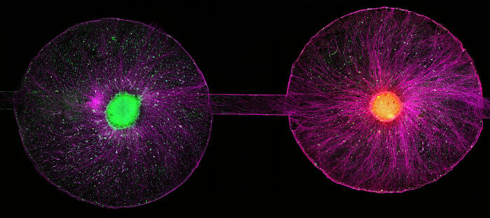
Hassanain Qambari, the first-place winner, shared: “The Nikon Small World competition is great, as it showcases amazing work across many disciplines from around the world. All the images presented in the competition represent the beauty and artistic side of science which may otherwise get overlooked. Such a competition not only celebrates the participants' hard work and passion but may also draw and inspire young scientists to pursue a career in STEM. It certainly inspired me.”
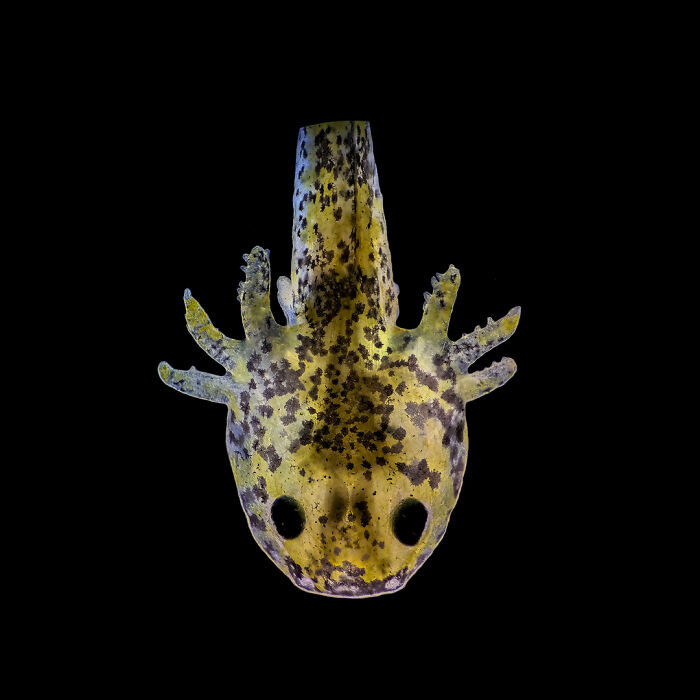
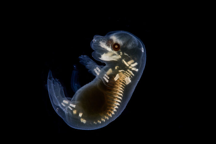
Universidad Nacional del ComahueDepartment of Biological SciencesSan Carlos de Bariloche, Río Negro, Argentina"One-week-old Axolotl after hatching."
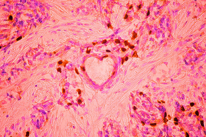
Independent Value-Based Healthcare Consultant Warsaw, Mazowieckie, Poland"Breast cancer cells."
Calling all scientists, photography and video enthusiasts! The Nikon Small World Competition, marking its 50th anniversary in 2024, invites everyone to join in. To be part of this incredible milestone, you can upload your digital images and videos directly onnikonsmallworld.com. Entry forms for Nikon’s 2024 Small World and Small World in Motion Competitions are available atenter.nikonsmallworld.com.
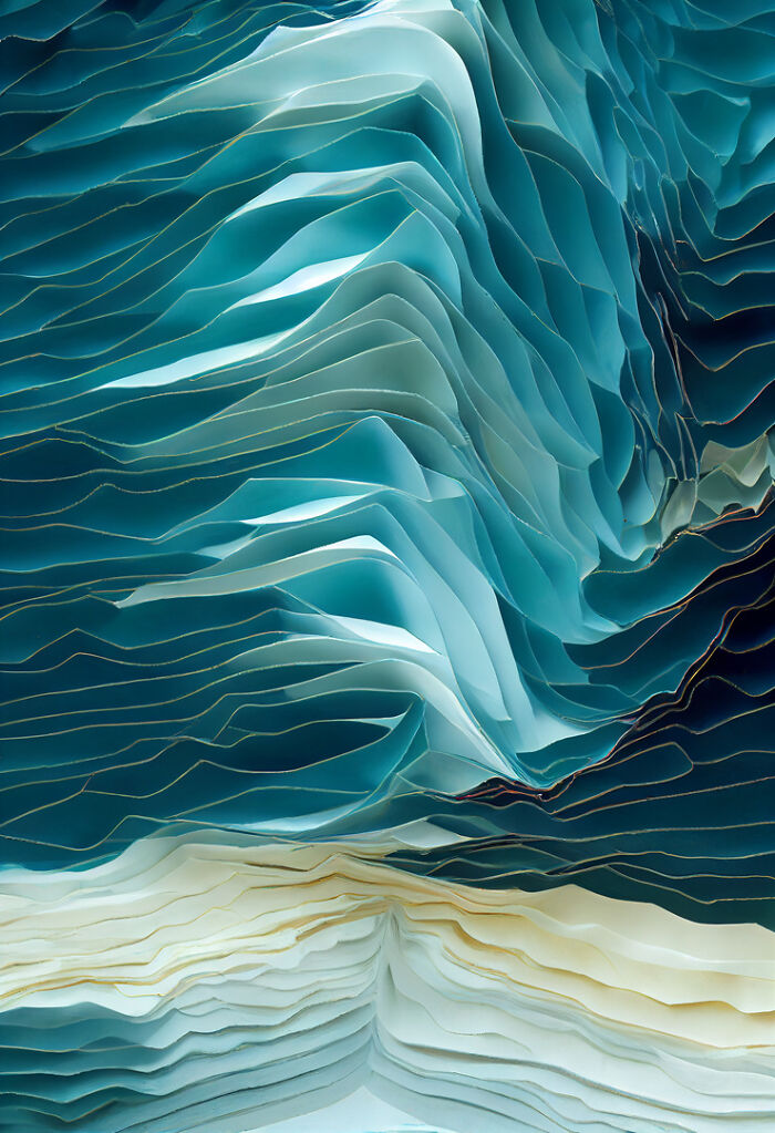
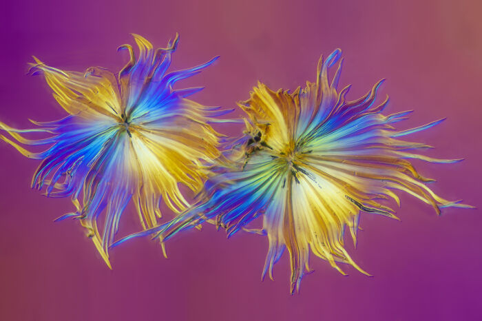
Impressions MicroscopiquesAmsterdam, Noord Holland, The Netherlands"Crystals of malonic acid dissolved in ethanol."
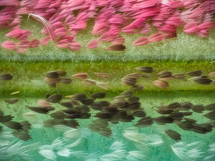
Rotterdam, Zuid-Holland, The Netherlands"Buckthorn trichomes."
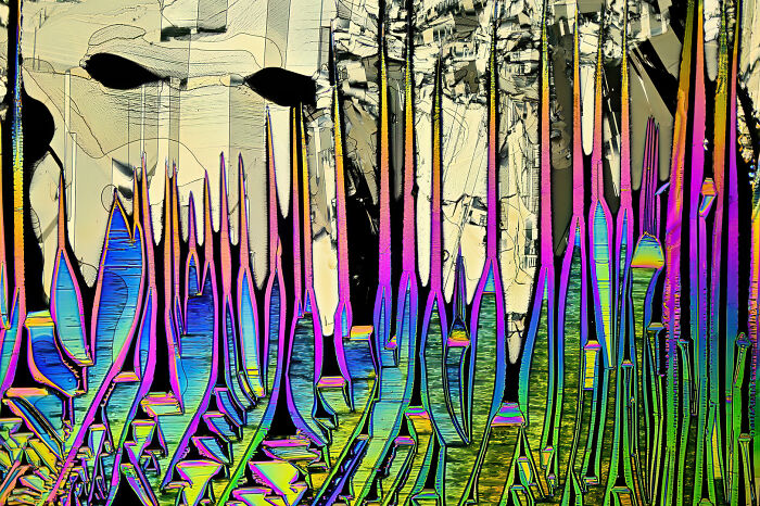
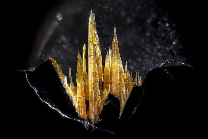
Mineralien LLCValley Village, California, USA"Golden rutile in quartz."
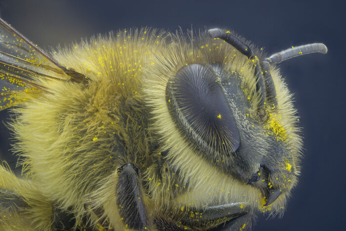
TCDD Teknik Müh. Müş. A.Ş.Ankara, Çankaya, Turkey"Bee"
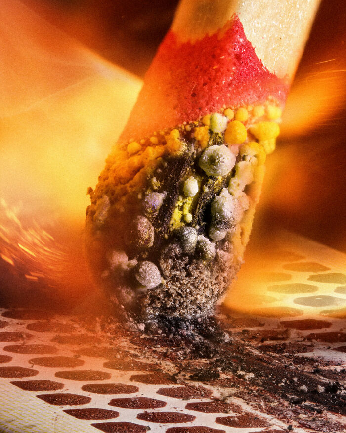
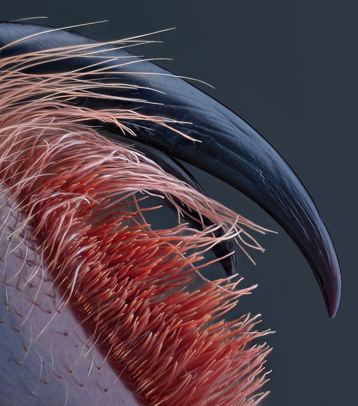
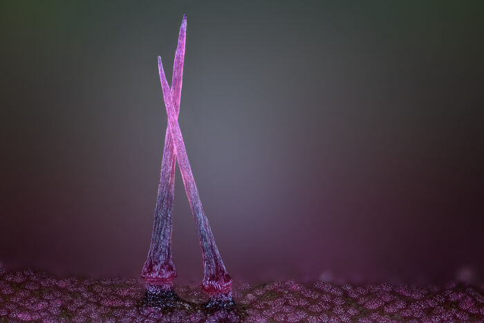
Medienbunker ProduktionBendorf, Rheinland Pfalz, Germany"Venomous fangs of a small tarantula."
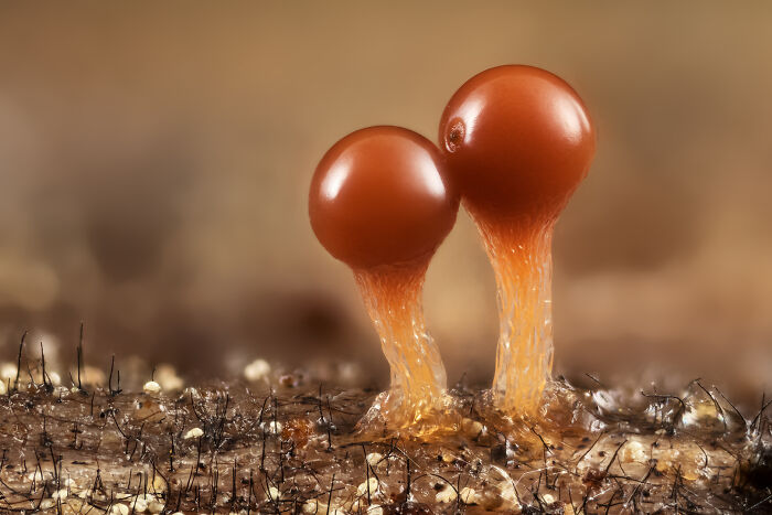
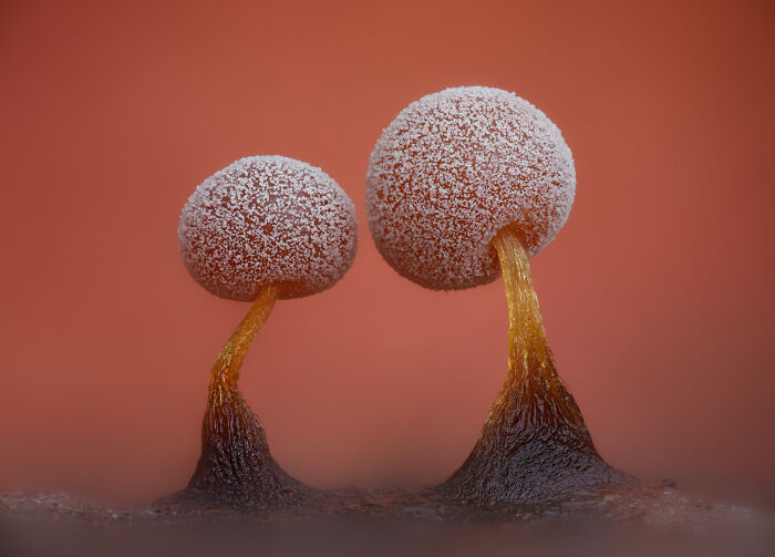
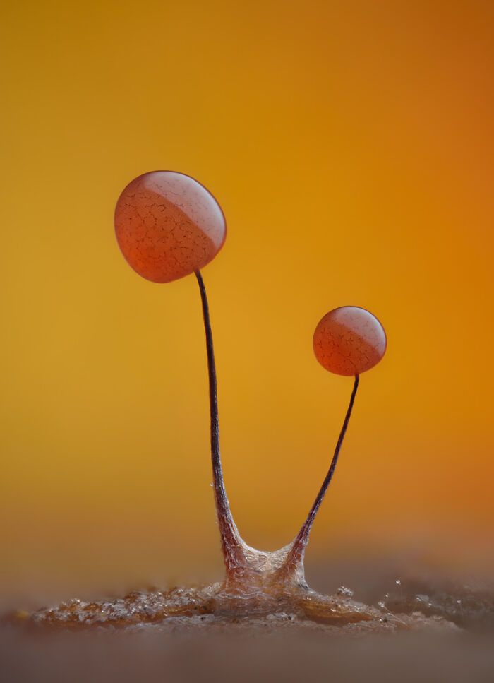
Svosov, Zilinsky, Slovakia"Slime mold (Trichia crateriformis)."
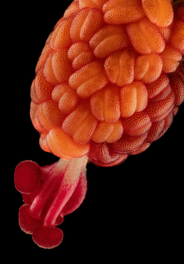
Raghuram Annadana PhotographyBangalore, Karnataka, India"Developing stamen and stigma inside a Hibiscus flower bud."
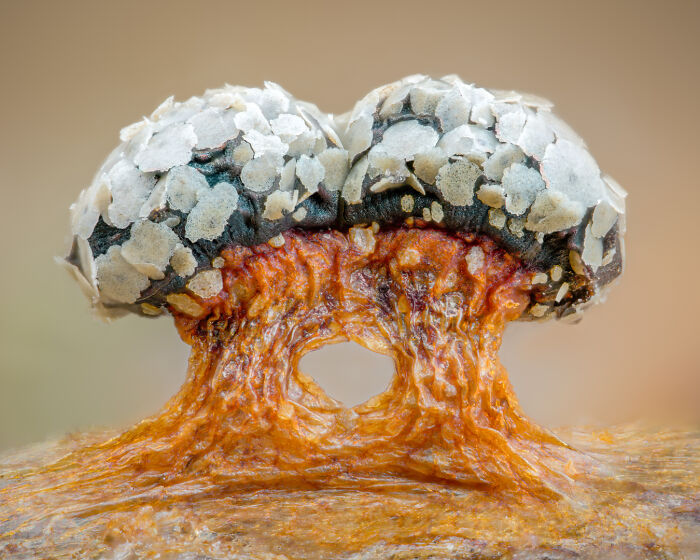
Charles Krebs PhotographyIssaquah, Washington, USA"Mushroom gills showing sporophores (sporangiophores)."
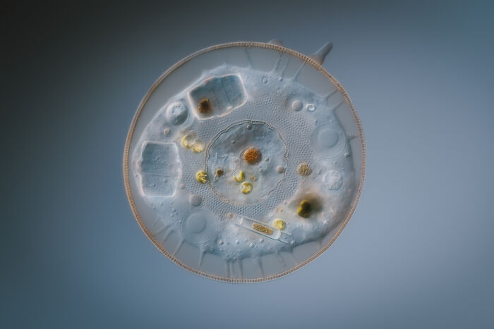
Bromma, Stockholm, Sweden"Amoeba (Arcella)."
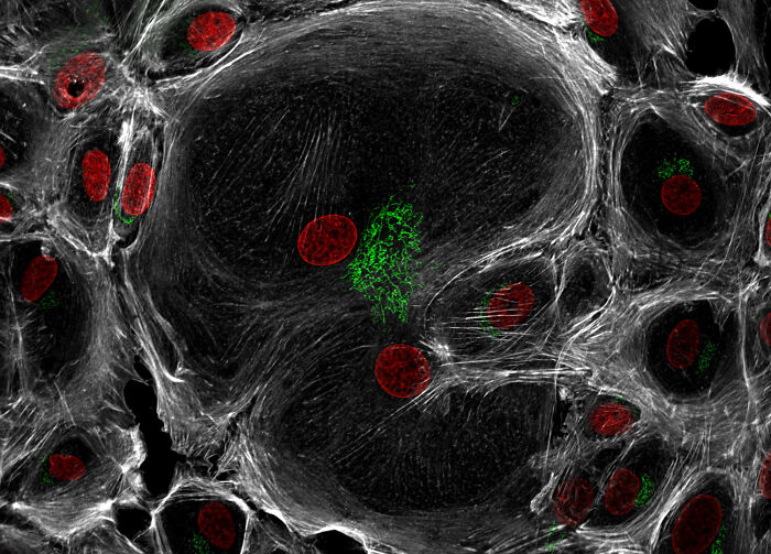
Medical University of South CarolinaDepartment of Regenerative Medicine & Cell BiologyCharleston, South Carolina, USA"Neonatal mouse intestinal tissue cells."
San Anselmo, California, USA"Slime mold (Diderma tigrinum)."
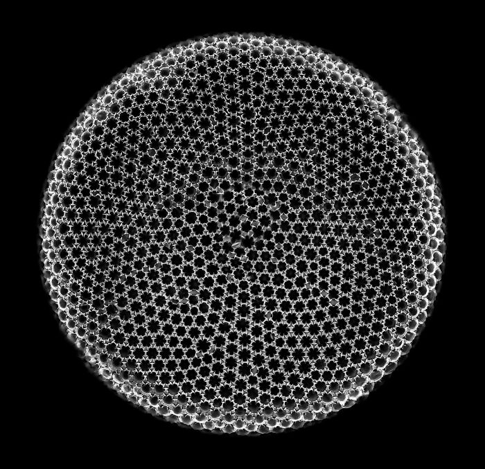
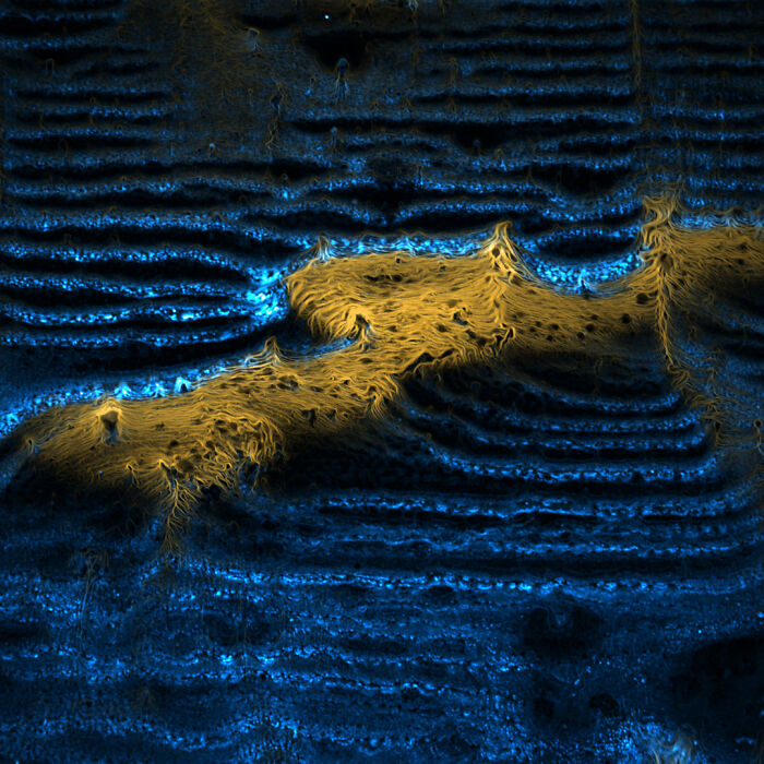
Weissensberg, Bavaria, Germany"Fossil diatom."
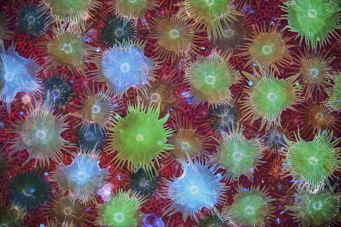
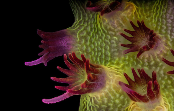
University of California, San DiegoScripps Institution of OceanographyLa Jolla, California, USA"Coral (Acropora granulosa) fluorescing under blue light."
WildMacroVacaville, California, USA"Slime mold (Didymium sp.) fruiting bodies."
Lions Eye InstituteDepartment of Physiology & PharmacologyPerth, Western Australia, Australia"Rodent optic nerve head showing astrocytes (yellow), contractile proteins (red) and retinal vasculature (green)."
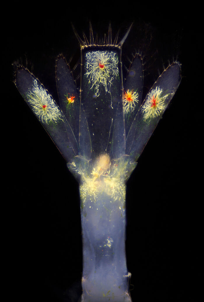
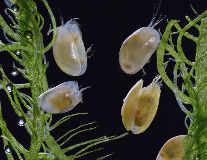
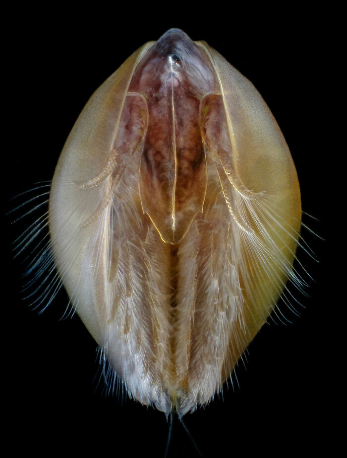
IES Virgen de la LuzDepartment of Biology and GeologyAvilés, Asturias, Spain"Tail of planktonic shrimp larvae."
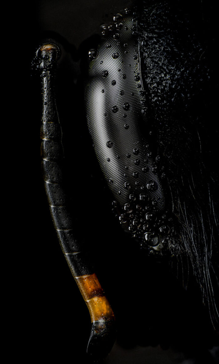
Madrid, Spain"Carpenter bee (Xylocopa violacea) head and antenna."
Feltwell, Norfolk, United Kingdom"Auto-fluorescing defensive hairs covering the leaf surface of Eleagnus angustifolia exposed to UV light."
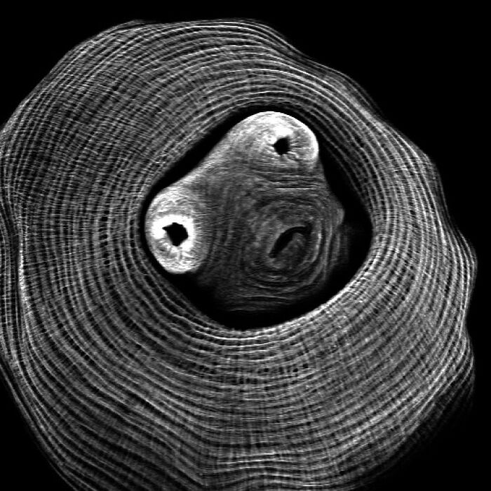
New Hope, Minnesota, USA"A cryptocrystalline micrometeorite resting on a #80 testing sieve."
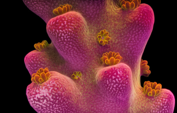
University of California, San DiegoScripps Institution of OceanographyLa Jolla, California, USA"Fluorescent image of an Acropora sp. showing individual polyps with symbiotic zooxanthellae."
University of HelsinkiIndividualized Drug Therapy Research Program, Faculty of MedicineHelsinki, Finland"Blood and lymphatic vasculatures in the ear skin of an adult mouse."
Continue reading with Bored Panda PremiumUnlimited contentAd-free browsingDark modeSubscribe nowAlready a subscriber?Sign In
Continue reading with Bored Panda Premium
Unlimited contentAd-free browsingDark mode
Unlimited content
Ad-free browsing
Dark mode
Subscribe nowAlready a subscriber?Sign In
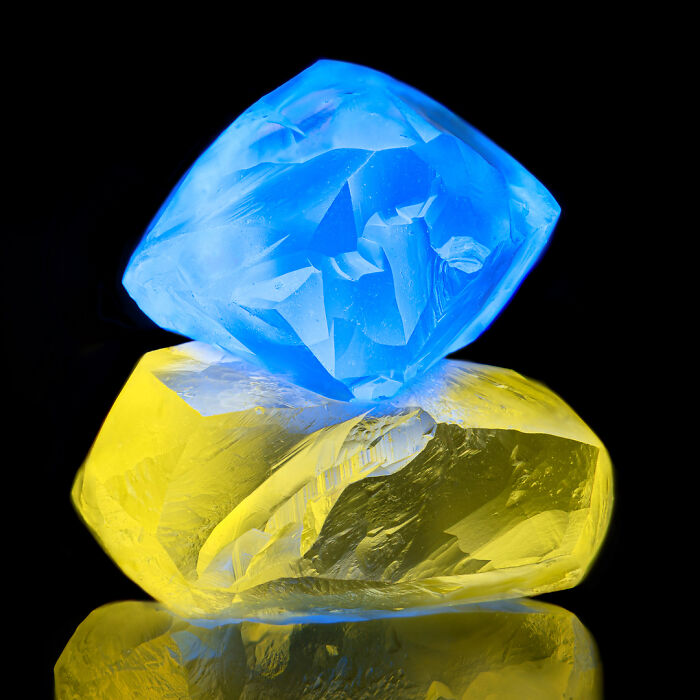
Don Komarechka PhotographyRavna Gora, Varna, Bulgaria"Two fluorescing diamonds."
Faculty of Science and Technology, Universiti Kebangsaan MalaysiaDepartment of Biology Science and BiotechnologyBangi, Selangot, Malaysia"Moss"
WildMacroVacaville, California, USA"Slime mold (Comatricha nigra) showing capillitial fibers through its translucent peridium."
Medienbunker ProduktionBendorf, Rheinland Pfalz, Germany"Cabbage butterfly eggs."
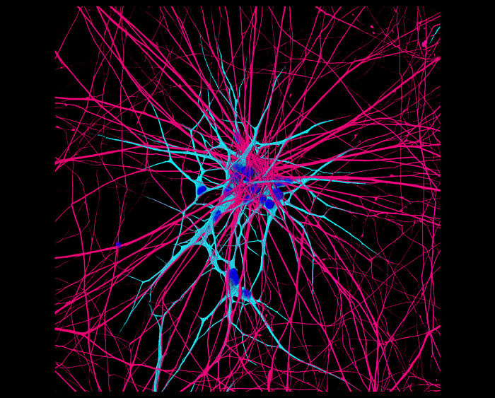
National Institutes of Health (NIH)NICHDBethesda, Maryland, USA"iPSC-derived human neurons."
Madrid, Spain"Mechanosensors in a Venus flytrap."
Nassau Community CollegeDepartment of BiologyGarden City, New York, USA"Ostracods and algae (Cladophora)."
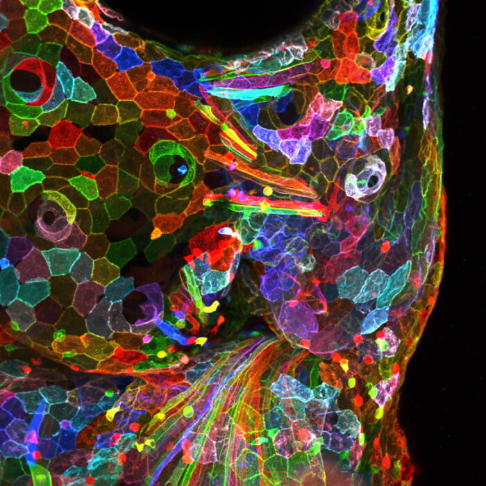
Academia SinicaChen-Hui Chen’s Lab in the Institute of Cellular and Organismic BiologyTaipei, Taiwan"Palmskin zebrafish larva."
Gaush Meditech Ltd.Hefei, Anhui Province, China"Bristle of a millipede (Polyxenidae)."
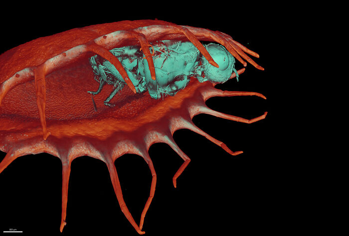
Calgara, Alberta, Canada"Clam shrimp (Lynceus mucronatus)."
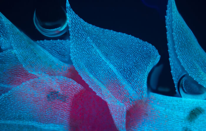
University of CambridgeSainsbury LaboratoryCambridge, Cambridgeshire, United Kingdom"Bindweed (Convolvulus) leaf epidermal cells autofluorescing."
University of GenevaDepartment of Genetics and EvolutionGeneva, Switzerland"Mouse embryo."

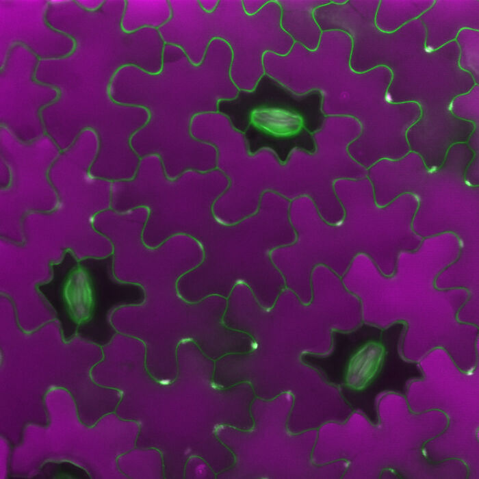
Marek Mis PhotographySuwalki, Podlaskie, Poland"Stomata in peace lily (Spathiphyllum sp.) leaf epidermis."
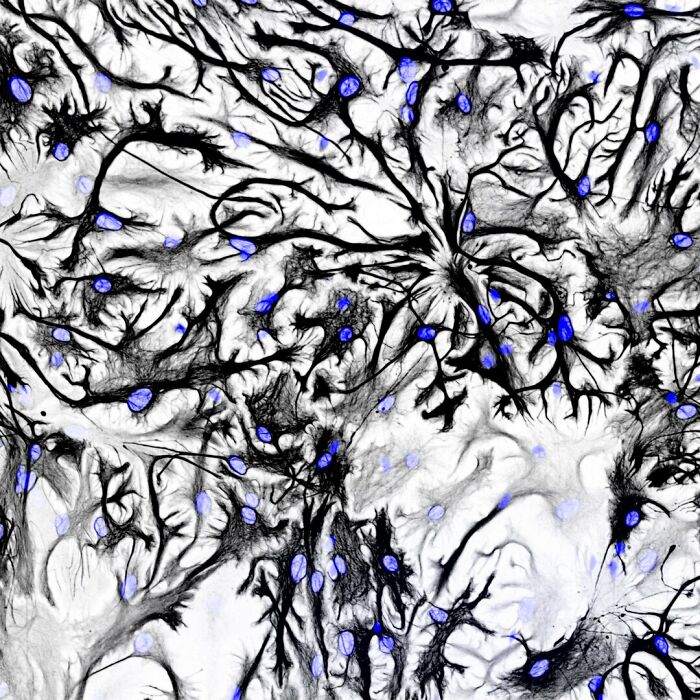
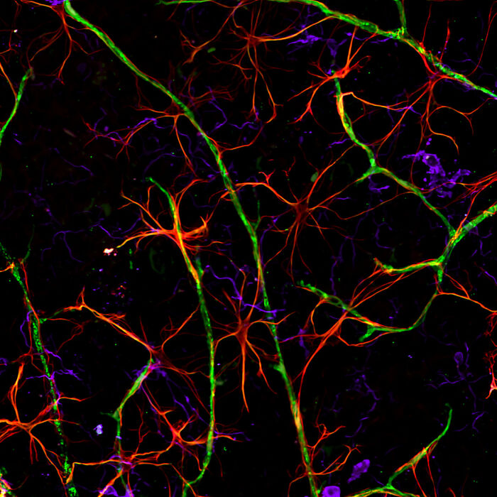
Freie Universität BerlinDepartment of BiochemistryBerlin, Germany"Rat astrocytes."
University of California, BerkeleyDepartment of Molecular and Cell BiologyBerkeley, California, USA"Algae from a mud puddle."
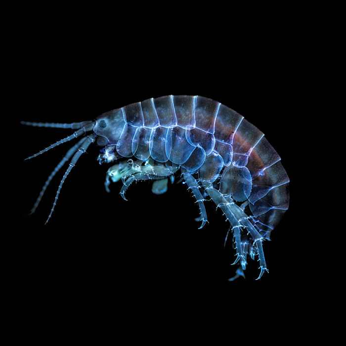
Gustometry + SF Micro SocietyNorwalk, Connecticut, USA"Freshwater amphipod."
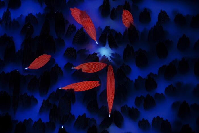
LaVision BioTec, a Miltenyi Biotec CompanyDepartment of BiophysicsBielefeld, North-Rhine Westphalia, Germany"Fly (cyan) caught in a Venus flytrap (red)."
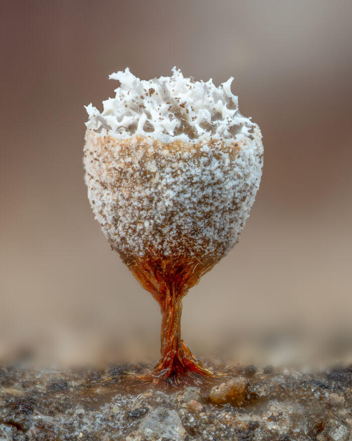
San Anselmo, California, USA"Slime mold (Craterium leucocephalum), looking like a beautiful tiny goblet."
See Also on Bored Panda
University of GenevaDepartment of Genetics and EvolutionGeneva, Switzerland"Dermal collagen in embryonic snake scales."
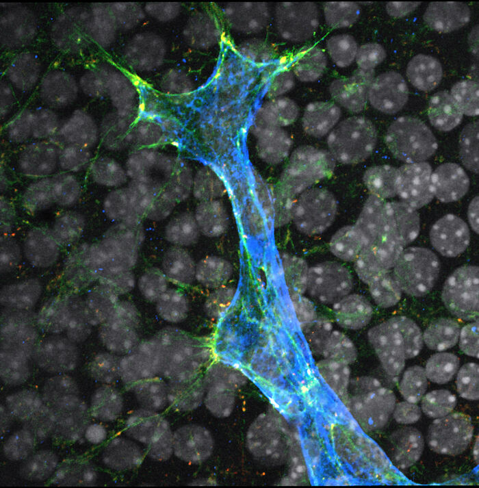
Macquarie UniversityMicroscopy FacilityMacquarie Park, New South Wales, Australia"Organ-on-chip system enabling the synaptic conjugation between 3D human embryonic stem cells."
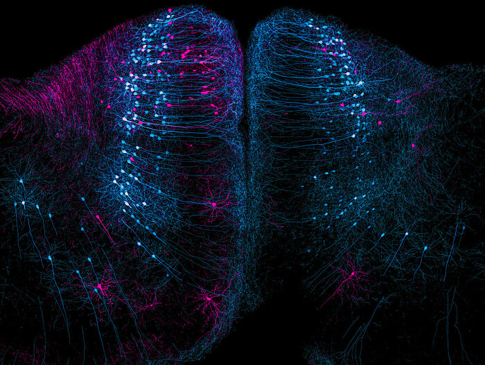
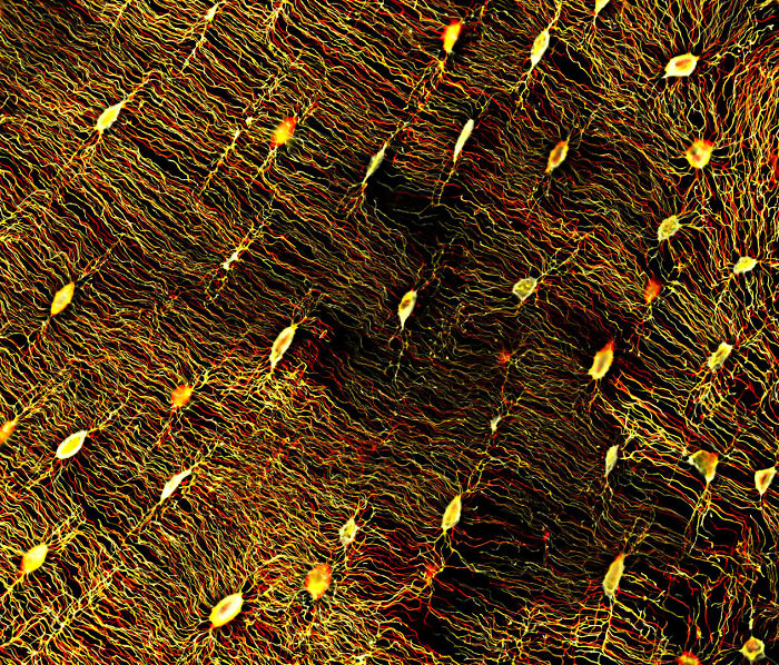
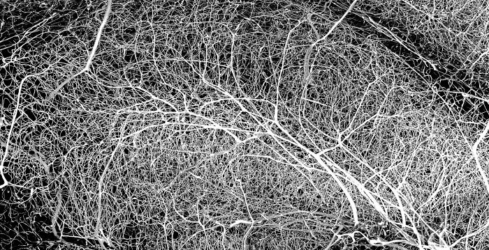
VIB (Flanders Institute of Biotechnology)Center for Brain and Disease ResearchLeuven, Vlaams-Brabant, Belgium"Retrograde labeled neurons in the cortex of a cortical mouse brain section."
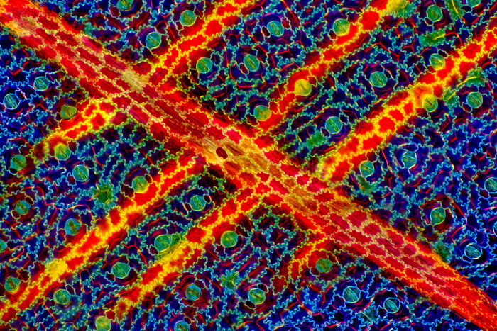
Universidad Complutense de MadridReal Sociedad Española de FísicaMadrid, Spain"Carbon nanotubes."
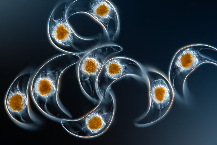
Trier University of Applied SciencesKonz, Rheinland-Pfalz, Germany"Marine organism (Pyrocystis lunula, Dinophyceae)."
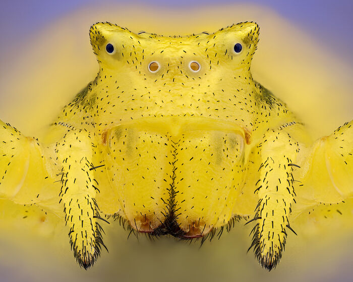
Saint Lys, Haute-Garonne, France"Crab spider (Thomisus onustus)."
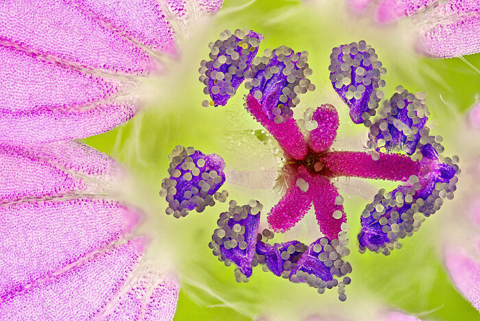
Saint Lys, Haute-Garonne, France"Geranium (Geraniaceae) stamen covered in pollen."
University of CambridgeSainsbury LaboratoryCambridge, Cambridgeshire, United Kingdom"Patterns at the surface of an embryonic leaf of Thale cress (Arabidopsis thaliana)."
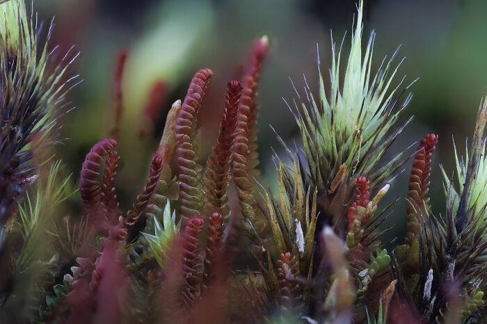
Rochester Institute of Technology (RIT)Department of Mechanical EngineeringRochester, New York, USA"Sphagnum moss with two air bubbles on the sample."
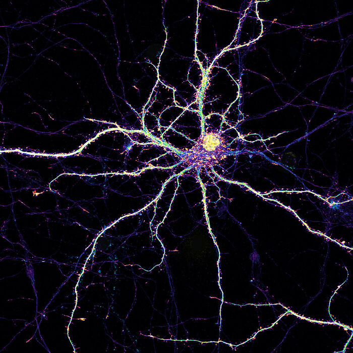
Amicus TherapeuticsPhiladelphia, Pennsylvania, USA"Maturing mouse cortical neuron in culture."
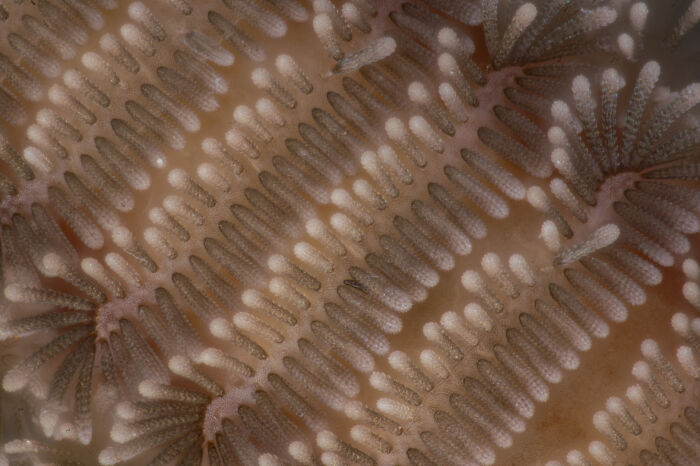
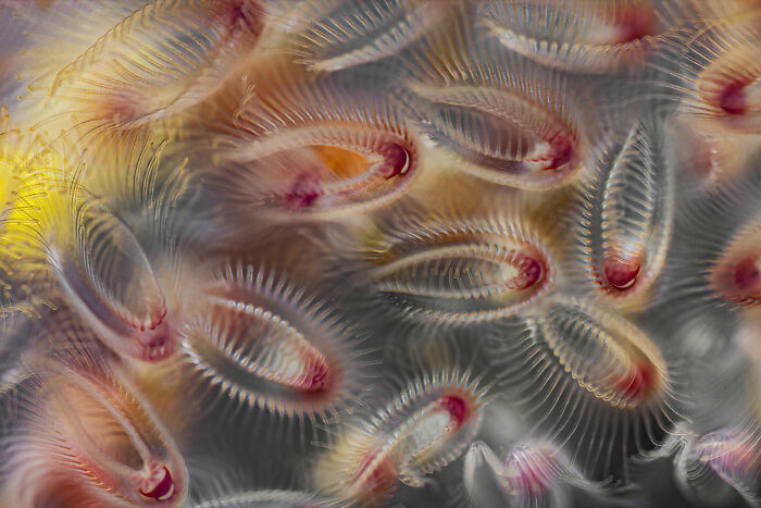
Charles Krebs PhotographyIssaquah, Washington, USA"Feeding bryozoan colony zooids. Bryozoans are microscopic aquatic invertebrates that live in colonies."
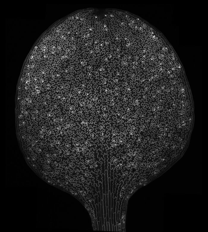
Northwestern UniversityBiological Imaging FacilityEvanston, Illinois, USA"Lily (Lilium) anther cross-section with pollen."
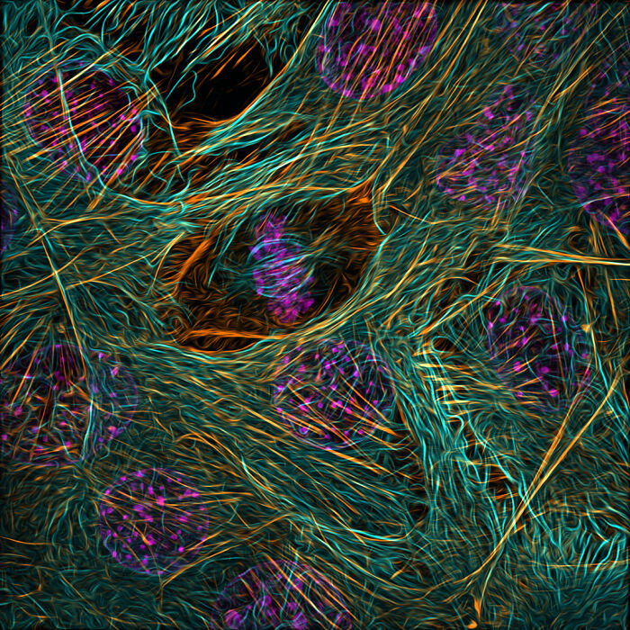
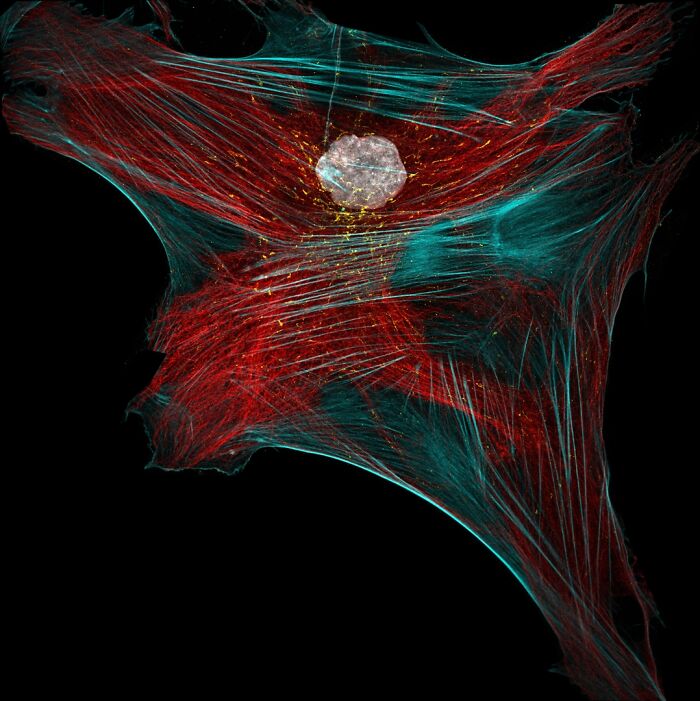
Baylor College of MedicineDepartment of Molecular Physiology and BiophysicsHouston, Texas, USA"Cytoskeleton of a dividing myoblast; tubulin (cyan), F-actin (orange) and nucleus (magenta)."
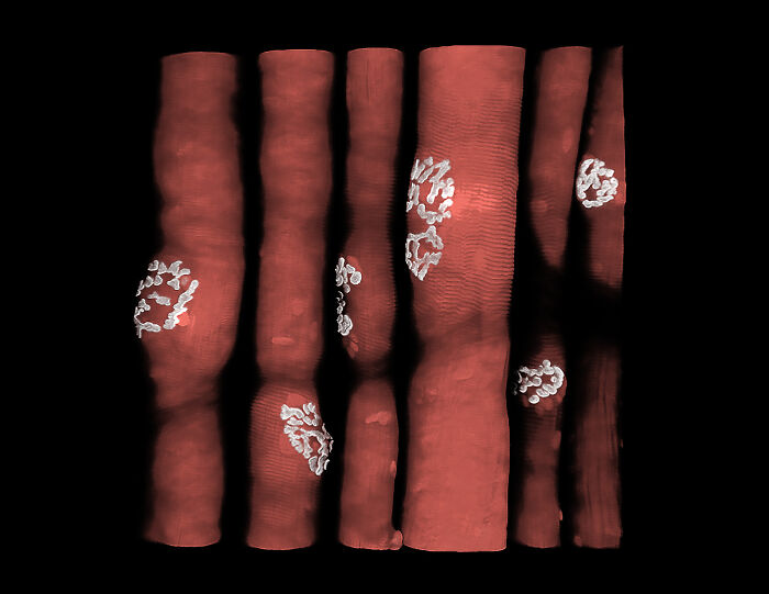
University of UtahHSC Cell Imaging CoreSalt Lake City, Utah, USA"Mouse femur bone lacunar-canalicular network (voids in bone that house osteocytes and their interconnected micro-tubular processes)."
Feltwell, Norfolk, United Kingdom"Wing scales of the cinnabar moth (Tyria jacobaeae) under ultraviolet light (UV)."
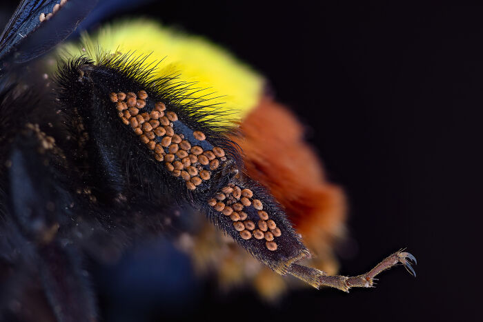
Higher Education Department Jammu and Kashmir IndiaDepartment of ZoologySrinagar, Jammu and Kashmir, India"Phoretic mites on the leg of a bumblebee."
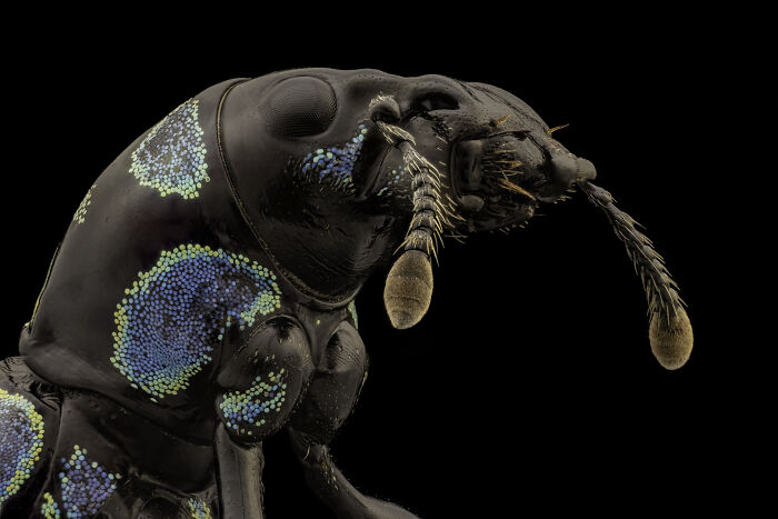
University of California, San Francisco (UCSF)Division of Transplant SurgerySan Francisco, California, USA"Blue black weevil (Metapocyrtus sp.)."
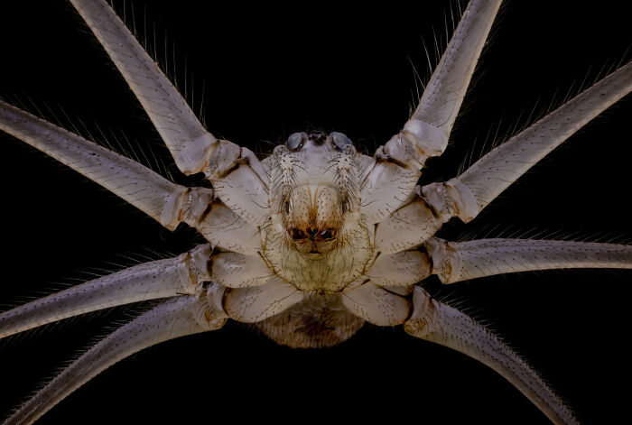
University of California, San Francisco (UCSF)Division of Transplant SurgerySan Francisco, California, USA"Underside of cellar spider (Pholcus phalangioides)."
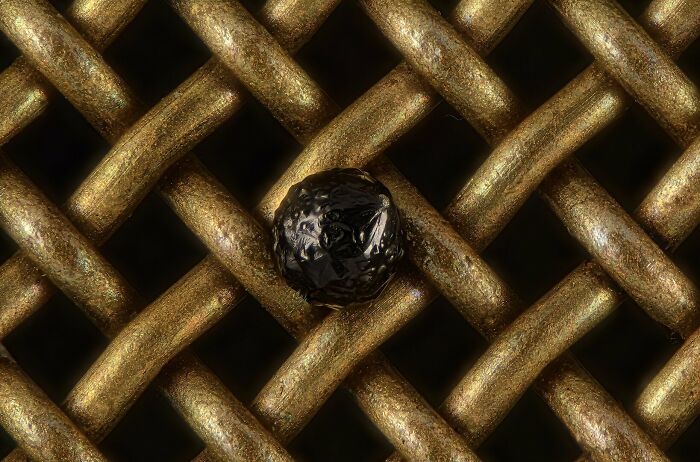
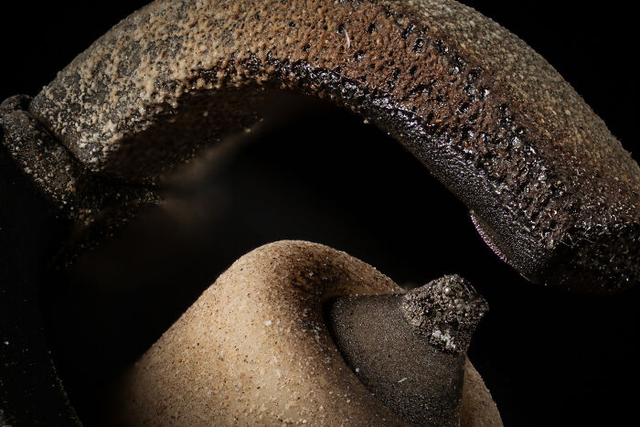
TeabeckVerona, Wisconsin, USA"Platinum spark plug."
National Autonomous University of Mexico (UNAM)Department of ImmunologyMéxico, Mexico"Muscle architecture of an evaginating tapeworm (Taenia crassiceps cysticercus)."
Macquarie UniversityMicroscopy FacilityMacquarie Park, New South Wales, Australia"Cleared mouse embryo."
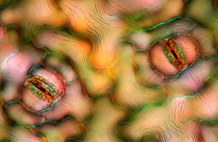
Marek Mis PhotographySuwalki, Podlaskie, Poland"Leaf epidermis stomata (Stromanthe sp.)."
University of KentuckySanders Brown Center on AgingLexington, Kentucky, USA"Mouse retina blood vessels (green) astrocytes (red) and microglia (purple)."
Institute of Biotechnology CASVestec, Central Bohemia, Czech Republic"Fluorescent actin filaments (yellow) and fluorescent anillin protein (blue) deposited on a glass coverslip after dewetting."
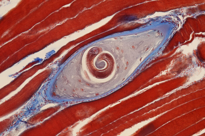
Modernist CuisineBellevue, Washington, USA"Trichinella cyst in pork muscle (Trichinella is a parasitic worm known to cause trichinosis)."
Academia SinicaChen-Hui Chen’s Lab in the Institute of Cellular and Organismic BiologyTaipei, Taiwan"In toto image of the skin and mucous cells in a live zebrafish larva."
Université du Québec à Trois-Rivières (UQTR)Department of Medical biologyTrois-Rivieres, Quebec, Canada"Distribution of cellular batteries (mitochondria-yellow) along the transport cables (Tubulin-red, actin-cyan) in human fibroblast."
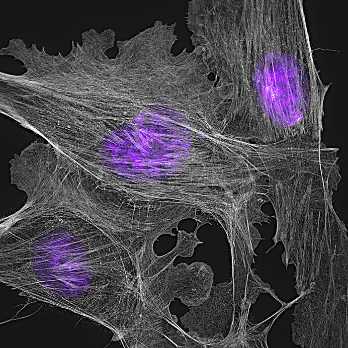
University of NottinghamSchool of Life Sciences Imaging (SLIM)Nottingham, USA"Actin cytoskeleton of bovine pulmonary epithelial cells."
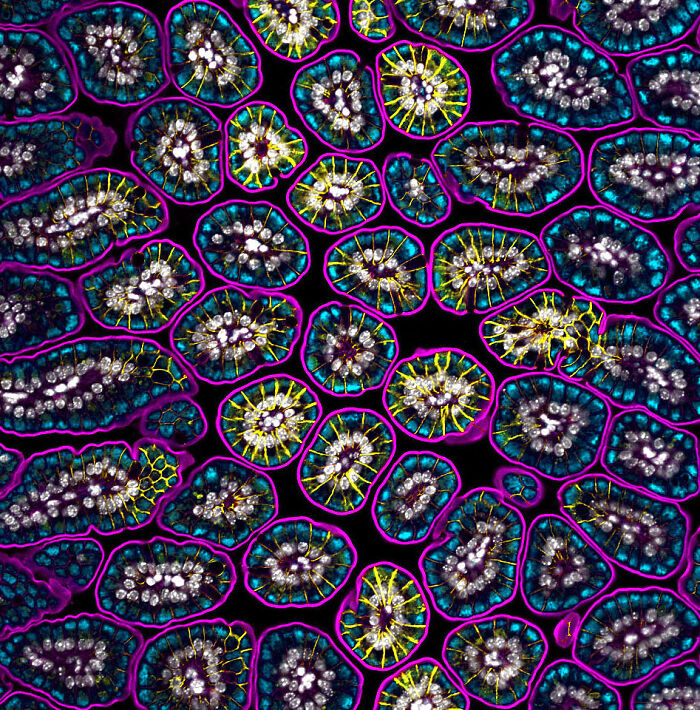
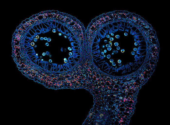
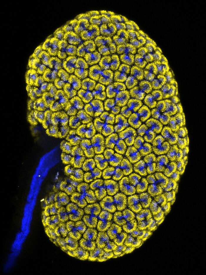
University of North Carolina at Chapel HillDepartment of Cell Biology and PhysiologyChapel Hill, North Carolina, USA"Embryonic mouse (Mus musculus) kidney showing the collecting duct (blue) and nephron progenitor (yellow) cells."
Rostock, Mecklenburg Vorpommern, Germany"Diatoms (single-celled algae) arranged on the head of a pin."
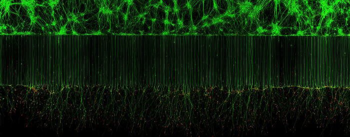
University of California, San DiegoDepartment of Cellular and Molecular MedicineLa Jolla, California, USA"Motor neurons grown in microfluidic device for separation of cell bodies (top) and axons (bottom). Green - microtubules; Red - growth cones (actin)."
University of MinnesotaDepartment of KinesiologySt. Paul, Minnesota, USA"Rat skeletal muscle fibers with associated neuromuscular junctions (white)."
Albert Einstein College of MedicineBronx, New York, USA"Non-parenchymal liver cells."
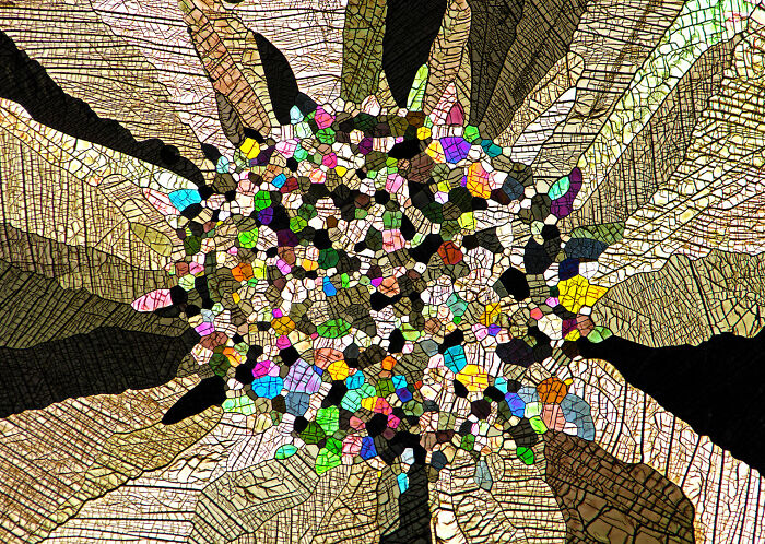
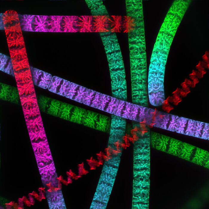
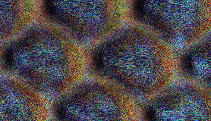
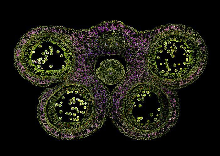
Ghent UniversityFaculty of Sciences/Faculty of Bioscience EngineeringGhent, Belgium"Composition of transverse sections of plant organs."
Uppsala UniversityDepartment of Immunology, Genetics and Pathology (IGP)Uppsala, Sweden"Blood and lymphatic vessels in a mouse diaphragm."
University of BordeauxBioTis-INSERM U1026Pessac, Gironde, France"Formation of blood vessels (angiogenesis) in the retina from a Lifeact-EGFP newborn mouse."
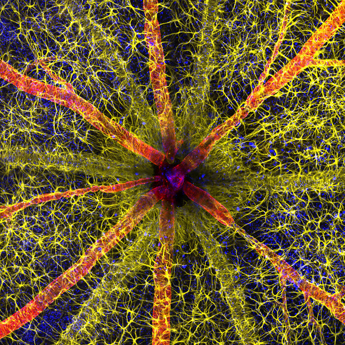
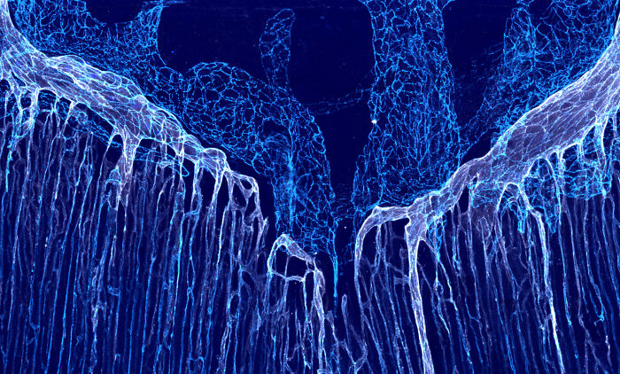
The Ohio State UniversityDepartment of NeuroscienceColumbus, Ohio, USA"3D capillary network section of the mammalian brain (dentate gyrus of the hippocampus)."
Modal closeAdd New ImageModal closeAdd Your Photo To This ListPlease use high-res photos without watermarksOoops! Your image is too large, maximum file size is 8 MB.Not your original work?Add sourcePublish
Modal close
Add New ImageModal closeAdd Your Photo To This ListPlease use high-res photos without watermarksOoops! Your image is too large, maximum file size is 8 MB.Not your original work?Add sourcePublish
Modal closeAdd Your Photo To This ListPlease use high-res photos without watermarksOoops! Your image is too large, maximum file size is 8 MB.Not your original work?Add sourcePublish
Add Your Photo To This ListPlease use high-res photos without watermarksOoops! Your image is too large, maximum file size is 8 MB.
Add Your Photo To This List
Please use high-res photos without watermarks
Ooops! Your image is too large, maximum file size is 8 MB.
Not your original work?Add source
Modal closeModal closeOoops! Your image is too large, maximum file size is 8 MB.UploadUploadError occurred when generating embed. Please check link and try again.TwitterRender conversationUse html versionGenerate not embedded versionAdd watermarkInstagramShow Image OnlyHide CaptionCropAdd watermarkFacebookShow Image OnlyAdd watermarkChangeSourceTitleUpdateAdd Image
Modal closeOoops! Your image is too large, maximum file size is 8 MB.UploadUploadError occurred when generating embed. Please check link and try again.TwitterRender conversationUse html versionGenerate not embedded versionAdd watermarkInstagramShow Image OnlyHide CaptionCropAdd watermarkFacebookShow Image OnlyAdd watermarkChangeSourceTitleUpdateAdd Image
Upload
UploadError occurred when generating embed. Please check link and try again.TwitterRender conversationUse html versionGenerate not embedded versionAdd watermarkInstagramShow Image OnlyHide CaptionCropAdd watermarkFacebookShow Image OnlyAdd watermark
Error occurred when generating embed. Please check link and try again.
TwitterRender conversationUse html versionGenerate not embedded versionAdd watermark
InstagramShow Image OnlyHide CaptionCropAdd watermark
FacebookShow Image OnlyAdd watermark
ChangeSourceTitle
You May Like40 Captivating Street Shots Curated By “Pure Street Photography"Community Panda86 Photography Hacks To Take Your Photos To The Next LevelAivaras KaziukonisPhotographer Assembles Aerial Photos To Showcase The Immense Scale Of Human Impact (17 Pics)Community Panda
Community Panda
Aivaras Kaziukonis
![]()
![]()
Photography
